- Bu konu 34 yanıt içerir, 3 izleyen vardır ve en son 14 yıl 10 ay önce
vet-34 tarafından güncellenmiştir.
-
YazarYazılar
-
8 Nisan 2009: 11:07 #75779
Murat KUTAY
Katılımcı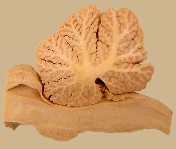
Sheep Hindbrain — Median View
The cerebellar vermis is sectioned sagittally in this median view of a sheep hindbrain. The vemis is divisible into about ten lobules; the most caudal lobule is the nodulus. Ventral to the cerebellum, the fourth ventricle is roofed by rostral and caudal medullary vellum. The latter gives rise to bilateral choroid plexuses.
8 Nisan 2009: 11:09 #75780Katılımcı
Canine Brain Illustration — Dorsal View
The vermis of the cerebellum is colored red in this dorsal view of an illustrated dog brain. The vermis is flanked bilaterally by cerebellar hemispheres
8 Nisan 2009: 11:10 #75781Katılımcı
Isolated Cerebellum — Ventral View
The cerebellum has been removed from the pons by cutting three cerebellar peduncles (white pics). The red pic in in the nodulus of the vermis. The orange pic is in the right flocculus. A flocculonodular tract (yellow) connects the two regions.
8 Nisan 2009: 11:13 #75782Katılımcı
Cranial Nerves — Canine Brain
Roots of the twelve cranial nerves are colored red (except olfactory nerves are not colored). Cranial nerves arise from the brainstem, except olfactory nerves which join the olfactory bulb. The trochlear nerve (IV) exits from the dorsal surface of the brainstem. Although it is regarded as a cranial nerve, the optic nerve (II) is embryologically and histologically an extension of the brain.
8 Nisan 2009: 11:15 #75783Katılımcı
Dissected Sheep Brain — Dorsal View
The top of the telencephalon has been removed to reveal lateral ventricles bilaterally. Notice that the floor of the lateral ventricle is formed by hippocampus (yellow), fimbria (green), and fornix (orange). Rostrally, the ventricle is bounded laterally by caudate nucleus (blue). (The red pic is in cerebral cortex and the white pic is in white matter (internal capsule).
8 Nisan 2009: 11:15 #75784Katılımcı
Lateral Ventricle in Sheep Cerebral Hemisphere
The hippocampus and fornix have been removed in this isolated cerebral hemisphere to show the lateral wall of the lateral ventricle. The caudate nucleus can be seen rostrally in the wall of the ventricle
8 Nisan 2009: 11:17 #75785Katılımcı
Dissected Equine Brain — Dorsal View
Ventricles are exposed in this dorsal view of a dissected equine brain. The lateral ventricle is the space medial to the caudate nucleus (1). The medial wall of the ventricle is thin between the corpus callosum and fornix. The third ventricle is evident anterior to the pineal body (green pic). The floor of the flourth ventricle (rhomboid fossa) is marked by white pics.
8 Nisan 2009: 11:18 #75786Katılımcı
Brain Ventricles — Dorsal & Lateral Views
Brain ventricles, including extensions into olfactory bulbs, are shown in dorsal (top) and lateral (bottom) views. An interventricular foramen connects each lateral ventricle with the third ventricle. A mesencepahlic aqueduct connects the third and fourth ventricles. Lateral recesses and appertures allow cerebrospinal fluid to exit to the subarachnoid space. Each ventricle has a choroid plexus that produces cerebrospinal fluid. Choroid plexuses are bilateral in third and fourth ventrticles.
8 Nisan 2009: 11:33 #75787Katılımcı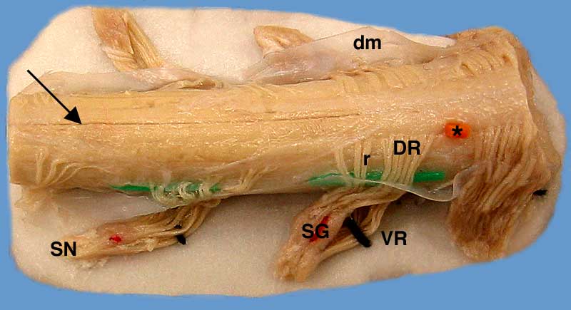
Spinal Cord Segments and Spinal RootsDural mater (dm) is reflected to expose segments and roots in a length of equine spinal cord. The arrow points to the dorsal median sulcus. The orange pic (asterisk) marks the dorsolateral sulcus, where dorsal roots enter the spinal cord. Each spinal cord segment gives rise to right and left dorsal and ventral spinal roots. Each spinal root is composed of rootlets (r). The dorsal root (DR) and the ventral root (VR) unite to form a spinal nerve (SN). A spinal ganglion (SG) is located distally on each dorsal root. Colored pics mark: spinal ganglia (red), the separation between dorsal and ventral roots (black), and the location of the denticulate ligament. The specimen rests on white cardboard.
8 Nisan 2009: 11:36 #75788Katılımcı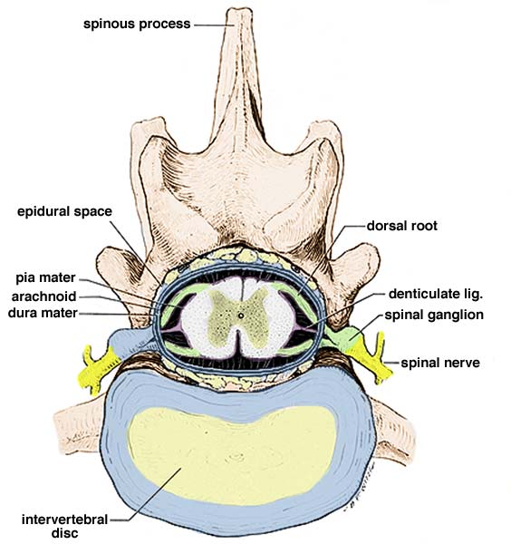
Spinal Cord Within Vertebral CanalThe following drawing depicts a spinal cord segment within a lumbar vertebra, at the level of an intervertebral disc (nucleus pulposus surrounded by annulus fibrosus). Spinal nerves are present bilaterally at intervertebral foramina, dorsal to the disc. An epidural space, containing fat, is evident external to spinal dura mater (blue). The latter is shown surrounding roots on the left; it is removed on the right side. Bilaterally, dorsal and ventral spinal roots (green) unite to form a spinal nerve (yellow) which soon branches. Bilateral thickenings of pia mater (purple), called denticulate ligaments, suspend the spinal cord within the dura mater.
8 Nisan 2009: 11:38 #75789Katılımcı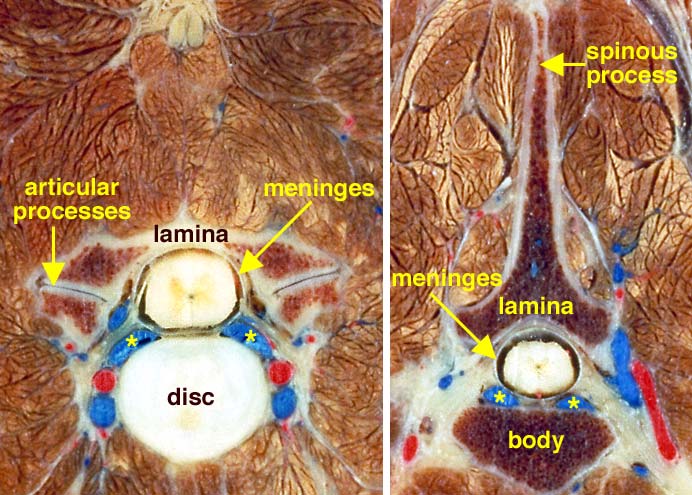
Spinal Cord In Situ
Left: Cervical transection through an intervertebral disc (nuchal ligament at the top). The spinal cord, surrounded by meninges, is evident within the vertebral canal. Internal vertebral venous sinus is marked by asterisks. (Vertebral a. & v. are visible bilaterally.)
Right: Thoracic vertebra transection. The spinal cord is surrounded by meninges within the vertebral canal. Internal vertebral venous sinus is marked by asterisks8 Nisan 2009: 11:39 #75790Katılımcı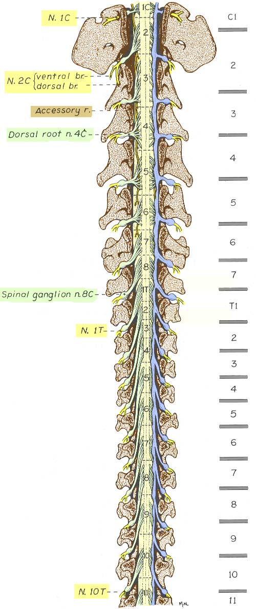
Canine Spinal Cord — Cranial HalfThe cranial half of a canine vertebral column has been drawn after a laminectomy to expose the spinal cord. Spinal cord segments are labeled and locations of vertebral bodies separated by intervertebral discs are labeled to the right.
Dura mater (blue) has been removed except along the right side. Dura mater envelops spinal roots including spinal ganglia.
Notice that spinal segments vary in length and that spinal roots must elongate to reach intervertebral foramina where segments are shifted cranially. In the cervical region, notice that the spinal root of the accessory cranial nerve (Accessory r., tan) emerges laterally, between dorsal and ventral roots. The C1 spinal nerve (N.1C, yellow) exits from a lateral foramen, rather than an intervertebral foramen like other spinal nerves. The C8 spinal segment appears to be “extra” (it lacks a nominally corresponding vertebra). Thus, caudal to the cervical region, spinal nerves exit through intervertebral foramina located at caudal margins of nominally corresponding vertebrae.
The C3 segment is the longest. Thereafter, segments progressively shorten in length. After the T2 segment, segments progressively lengthen. The cervical enlargement (C6, 7, 8, & T1) which innervates the thoracic limb (brachial plexus) is centered approximately at the C6-C7 intervertebral disc.
8 Nisan 2009: 11:40 #75791Katılımcı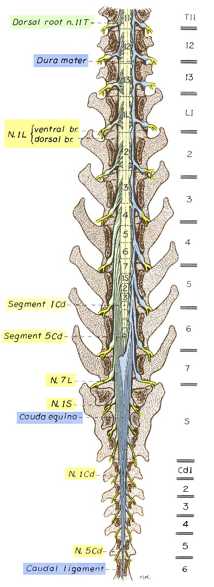
Canine Spinal Cord — Caudal HalfThe cranial half of a canine vertebral column has been drawn after a laminectomy to expose the spinal cord. Spinal cord segments are labeled and locations of vertebral bodies separated by intervertebral discs are labeled to the right.
Dura mater (blue) has been removed except along the right side. Dura mater envelops spinal roots including spinal ganglia.
Notice that thoracolumbar spinal segments are long and located within nominally corresponding vertebrae. Thereafter, segments progressively shorten in length and spinal roots elongate as segments shift position cranial to nominally corresponding vertebrae. Sacral and caudal roots streaming caudally are referred to as the cauda equina. Notice that the cauda equina is initially intrathecal (within the main cylinder of spinal dura mater); thereafter, the roots are enveloped by dural sheaths in the epidural space.
The term conus medullaris refers to the cone-shaped region of spinal cord caudal to the lumbosacral enlargement (L4 — S1). The cord terminates approximately at the L6-L7 intervertebral disc. Thereafter a terminal filament of glial tissue continues for some distance. The term caudal ligament refers to the terminal filament enveloped by a dural sheath.
8 Nisan 2009: 11:41 #75792Katılımcı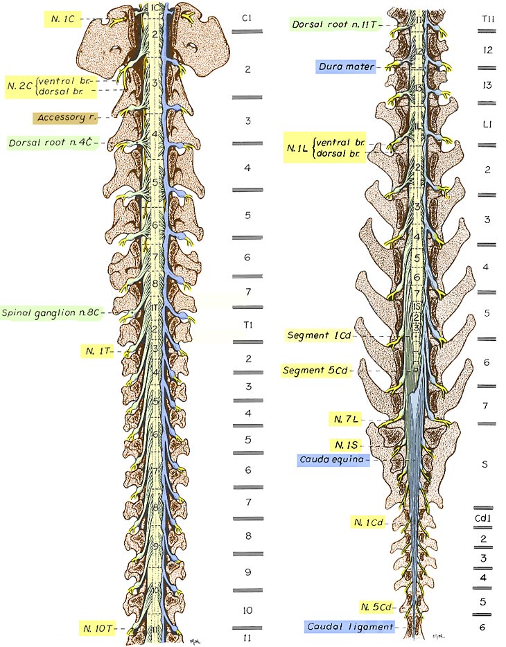
Canine Spinal CordCranial and caudal halves of a canine vertebral column are illustrated, after a laminectomy to expose the spinal cord. Spinal cord segments are labeled, and locations of vertebral bodies separated by intervertebral discs are shown to the right. Dura mater (blue) has been removed except along the right side. The illustrated position relationship of spinal cord segments to vertebrae represents the most common relationship for medium and large dogs (typical variation is half a vertebral length cranial or caudal to that shown). In small dogs (under 7kg) spinal cord segments are positioned more caudally than is shown.
8 Nisan 2009: 11:42 #75793Katılımcı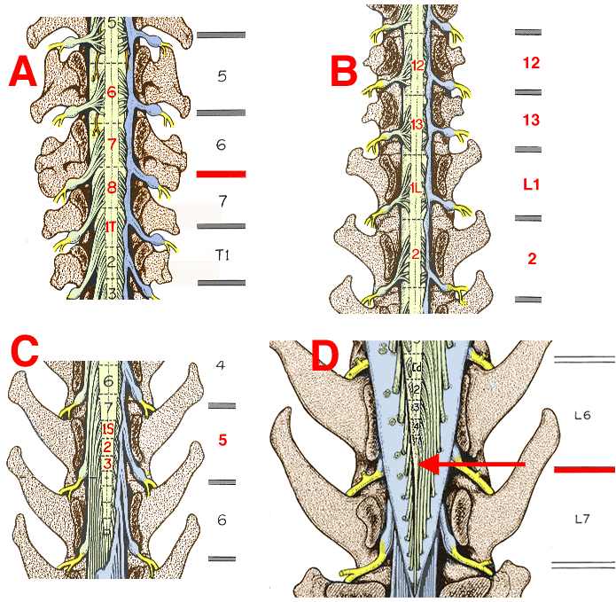
Spinal Cord—Vertebrae RelationshipsIt is clinically useful to know the approximate locations of spinal cord segments relative to palpable, radiographically visible vertebrae. One learning strategy is to remember the following four relationships and then interpolate other position relationships as necessary. (The illustrated relationships are the most common for medium and large dogs (± half vertebra). In small dogs the position is one vertebra caudal to that shown.)
A. The cervical enlargement (brachial plexus segments) are centered at the C6-C7 intervertebral disc.
B. At the thoraco-lumbar junction, segments are positioned within nominally corresponding vertebrae.
C. The three sacral segments are located within the L5 vertebra.
D. The spinal cord terminates at the L6-L7 intervertebral disc. -
YazarYazılar
- Bu konuyu yanıtlamak için giriş yapmış olmalısınız.